You have no items in your shopping cart
Prepared slides set 23-PC
- Buy 2 and save 5%
Euromex offers both teachers, students and microscopy amateurs a wide range of quality microslides, which can be used immediately in biology classes as introduction to zoology and botany
23 Slides sets of human and mammal histology
(for education)
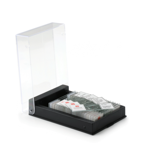
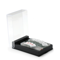
Set with 23 microscopy slides for education: human histological samples of men and mammals. Each set contains a variety of prepared slides (see 'Specification').
All microscope slides supplied in a plastic box with transparant cover for 25 slides
Highlights
- 23-PC Prepared slides set
- High quality microscope slides
- Human and mammal histology
- Set for education
$$$$$
How Are Cells Stained and Slides Prepared?
Cell staining techniques and preparation depend on the type of stain and analysis used. One or more of the following procedures may be required to prepare a sample:
• Fixation - serves to "fix" or preserve cell or tissue morphology through the preparation process. This process may involve several steps, but most fixation procedures involve adding a chemical fixative that creates chemical bonds between proteins to increase their rigidity. Common fixatives include formaldehyde, ethanol, methanol, and/or picric acid.
• Mounting - involves attaching samples to a glass microscope slide for observation and analysis. Cells may either be grown directly to the slide or loose cells can be applied to a slide using a sterile technique. Thin sections (slices) of material such as tissue may also be applied to a microscope slide for observation.
–––––––––––––––––––––––––––––––––––––––––––––––––––––––––
PB.5221 23 Set of human and mammal slides (education)
The set contains:
EPHITELIUM AND CONNECTIVE TISSUES
SH.1001 Loose connective tissue, rabbit
SH.1005 Hyaline cartilage rabbit, section
SH.1011 Hard bone grinding, human, section
SH.1049 Muscle types, striated, smooth, heart muscle, dog, l.s.
SH.1060 Tendon, dog, l.s.
SH.1070 Squamous epithelium, human, isolated cells from mouth, smear
SH.1072 Skin section through hair follicle, human
SH.1078 Stratified flat epithelium, dog, section
RESPIRATORY, BLOOD CIRCULATION AND ENDOCRINE SYSTEM
SH.1120 Trachea rabbit, c.s.
SH.1130 Artery and vein, rabbit, c.s.
SH.1150 Blood smear human, Giemsa stained
DIGESTIVE SYSTEM
SH.1210 Stomach wall, dog, section
SH.1230 Small intestine, dog, c.s.
SH.1250 Liver, pig, section
URINARY AND GENITAL SYSTEM
SH.1310 Injected kidney rabbit, sec.
SH.1330 Testis rabbit, c.s.
SH.1340 Ovary rabbit with developed eggs
SH.1376 Human chromosomes in blood, female
NERVOUS SYSTEM AND SENSORY ORGANS
SH.1410 Nerve, rabbit, c.s. and l.s.
SH.1430 Cerebrum section, dog
SH.1450 Spinal cord, rabbit, c.s.
SH.1470 Taste buds, rabbit, l.s.
SH.1490 Retina, rabbit, sec.
–––––––––––––––––––––––––––––––––––––––––––––––––––––––––
Used abbreviations:
c.s. = cross section
l.s. = longitudinal section
w.m. = whole mount
–––––––––––––––––––––––––––––––––––––––––––––––––––––––––
PB.5222 23 Set of human and mammal slides (education)
The set contains:
EPHITELIUM AND CONNECTIVE TISSUES
SH.1006 Elastic cartilage, rabbit
SH.1012 Hard bone grinding of rabbit, tooth, de-calcium
SH.1040 Smooth muscle, teased preparation, rabbit, l.s.+c.s.
SH.1043 Cardiac muscle, dog, l.s.
SH.1045 Skeletal muscle, dog, l.s and c.s.
SH.1075 Skin section with sweat gland, human
SH.1080 Ciliated epithelium, trachea, rabbit
RESPIRATORY, BLOOD CIRCULATION AND ENDOCRINE SYSTEM
SH.1110 Lung with injected blood vessels, rabbit, c.s.
SH.1140 Pancreas, rabbit, section
SH.1160 Lymph node, rabbit, section
SH.1170 Thyroid gland, rabbit, section
SH.1180 Adrenal gland, rabbit, section
DIGESTIVE SYSTEM
SH.1220 Oesophagus dog, c.s.
SH.1235 Large intestine, dog, c.s.
SH.1260 Gall bladder, dog, section
SH.1280 Golgi apparatus in Basal spinal ganglion, dog
URINARY AND GENITAL SYSTEM
SH.1315 Kidney rat, sec. cortex & medulla
SH.1360 Sperm human, smear
SH.1375 Human chromosomes in blood, male
SZ.1724 Fruitfly, drosophila, giant chromosomes of salivary gland, w.m.
NERVOUS SYSTEM AN SENSORY ORGANS
SH.1415 Motor nerve cells with end plates, rabbit, w.m.
SH.1420 Cerebellum section, dog
SH.1480 Eyeball section, rabbit, sagittal section
–––––––––––––––––––––––––––––––––––––––––––––––––––––––––
Used abbreviations:
c.s. = cross section
l.s. = longitudinal section
w.m. = whole mount
–––––––––––––––––––––––––––––––––––––––––––––––––––––––––

